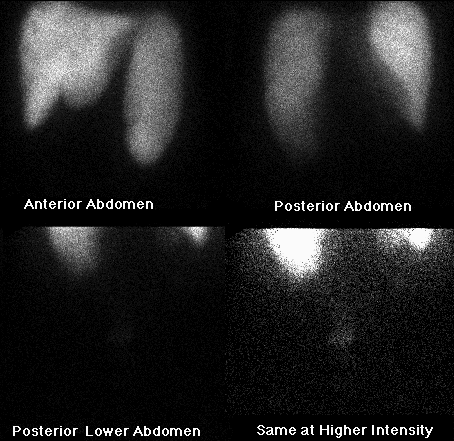
DELAYED SPOT VIEWS OF THE UPPER AND LOWER ABDOMEN
View main image(bm) in a separate image viewer
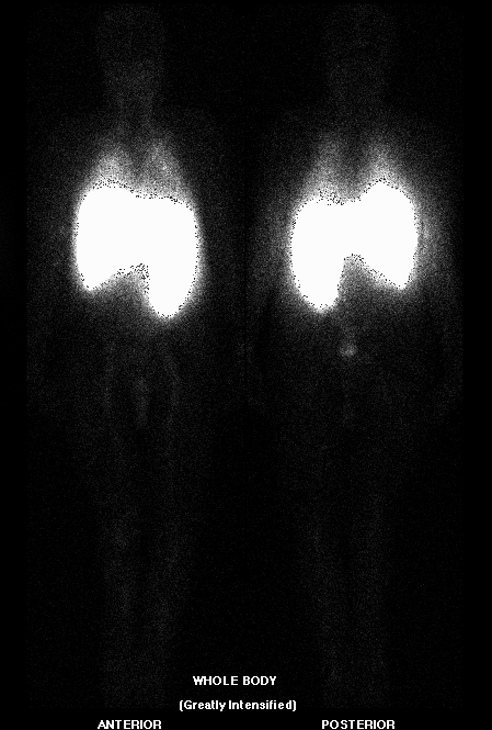
View second image(bm). WHOLE BODY ANTERIOR AND POSTERIOR IMAGES (GREATLY INTENSIFIED)
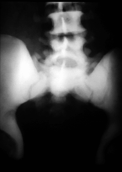
View third image(xr). AP VIEW OF THE PELVIS
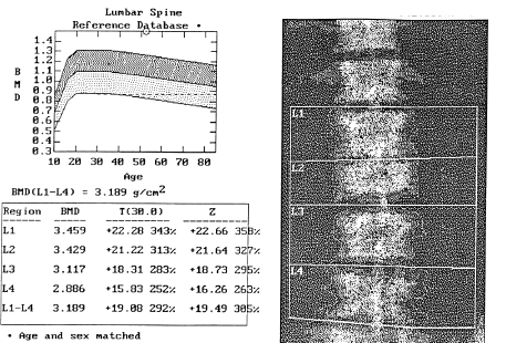
View fourth image(mm). DUAL ENERGY XRAY ABSORPTIOMETRY OF LUMBAR SPINE
Full history/Diagnosis is available below
There is massive splenomegaly--likely representing the principal site of extramedullary hematopoeisis. Slightly less activity is present in the spleen as compared with the liver.
Plain film of the pelvis reveals diffuse dense bony sclerosis consistent with the patient's known osteopetrosis.
Dual energy xray absorptiometry of the lumbar spine revealed an average bone mineral density of 3.19 g/cm2 in this patient. This bone density is 19.5 standard deviations higher than the normal value in a male patient of the same age!
Bone marrow scintigraphy is seldom used today but can be helpful in certain clinical situations, such as with this patient. The procedure involves the intravenous injection of small (pre-filtered) colloid particles (0.1-0.22 um) which are taken up by the reticuloendothelial system (RES). Normally, Tc99m sulfur colloid (which has not been filtered and thus includes particles as large as 0.5 um) localizes predominanatly in the Kupffer cells of the liver (85%), macrophages of the spleen (10%), and bone marrow (5%).
Factors which influence to distribution include extraction efficiency, blood flow, disease states, and particle size. Larger particles localize within the liver, while smaller particles collect in the bone marrow.
In most normal subjects, red marrow is seen in the axial skeleton and proximal appendicular skeleton (proximal thirds of the femur and humeri). In osteopetrosis one often sees decreased central marrow uptake with peripheral expansion (increased marrow in the distal extremities) and/or extramedullary hematopoiesis (ie. hepatosplenomegaly).
This patient does not demonstrate peripheral marrow expansion, but does have an enlarged functioning spleen showing increased uptake due to increased number and activity of spenic macrophages.
This study provided useful information in this case. We determined that this patient's situation is tenuous because the spleen has become the primary site of hematopeoisis, while the enlargement of the organ has led to platelet sequestration and subsequent thrombocytopenia. The tentatively scheduled splenectomy was thus cancelled.
References and General Discussion of Bone Marrow Scintigraphy (Anatomic field:Skeletal System, Category:Other generalized systemic disorder)
Return to the Teaching File home page.