Case Author(s): Samuel Wang, M.D. and Barry A. Siegel, M.D. , 5/18/97 . Rating: #D3, #Q4
Diagnosis: Bone infarctions.
Brief history:
18-year-old male with
sickle cell anemia and left leg pain.
Images:

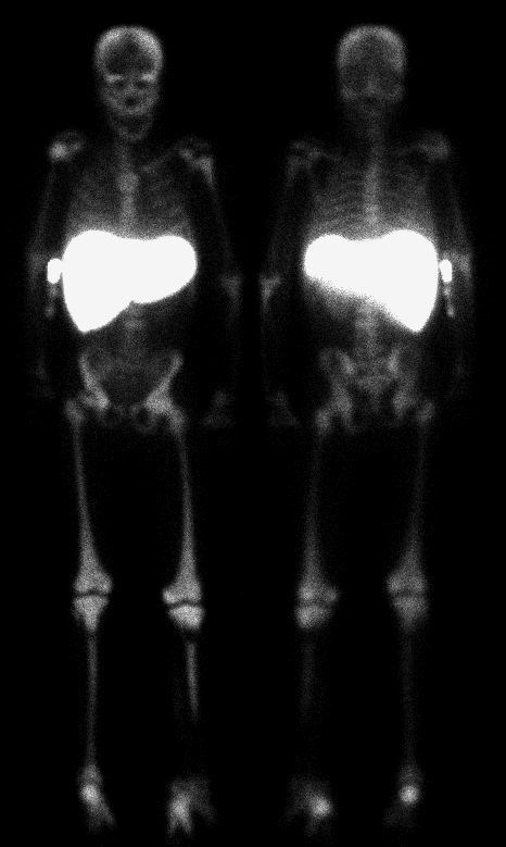
Delayed 18-hour anterior and posterior whole-body images.
View main image(iw) in a separate image viewer
View second image(bm).
Anterior and posterior whole-body images after injection of a second radiopharmaceutical.
View third image(bm).
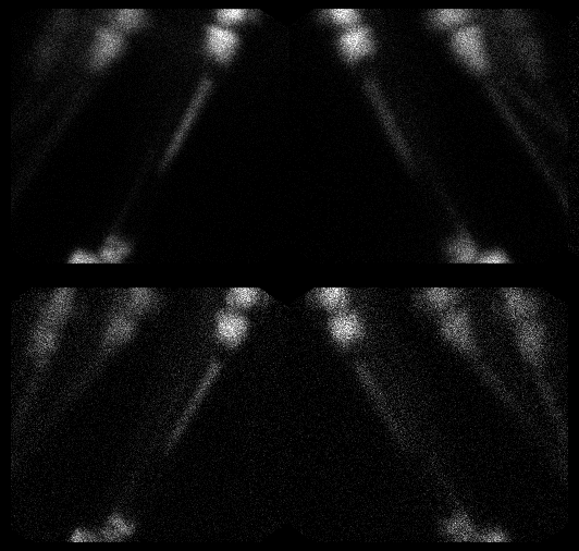
Spot views comparing the distribution of the two radiopharmaceuticals
Full history/Diagnosis is available below
Diagnosis: Bone infarctions.
Full history:
18-year-old male with a
history of sickle cell anemia (hemoglobin SS
disease) who presented with a one-week history
of pain in the left tibia. The patient's white
blood cell count was 28,000/µL, the ESR was 60
mm/hr, and he had fevers. The patient was
referred to help differentiate osteomyelitis from
bone infarction. Approximately 18 hours after
injection of In-111 labeled leukocytes, anterior
and posterior whole-body images (main image)
were obtained along with additional spot
images of the left leg (third image, 2nd row). After
In-111 imaging of the left leg was complete,
imaging of the marrow distribution in the left
leg was performed by injecting Tc-99m sulfur
colloid (third image, 1st row). The patient was not moved between
the In-111 WBC imaging and the Tc-99m SC
imaging of the left leg. Finally, anterior and
posterior whole-body Tc-99m sulfur colloid
images were obtained (second image).
Radiopharmaceutical:
0.45 mCi In-111
labeled autologous leukocytes i.v. and 10.1 mCi
Tc-99m sulfur colloid i.v.
Findings:
There are no significant
differences in the distribution of marrow
activity on the In-111 WBC images and the Tc-
99m SC images. Specifically, on both sets of
images there are focal areas of absent activity
seen in the left proximal tibia and in the distal
third of the left tibia. Normal marrow activity
is seen with both radiopharmaceuticals in the
mid left tibia. These findings are not consistent
with osteomyelitis of the tibia and more likely
represent infarcts of the proximal and distal
portions of the left tibia.
Both the In-111 WBC and the Tc-99m SC images
demonstrate absent activity in the right clavicle
and left humeral head, compatible with
previous infarctions. Heterogeneous activity in
the lower lumbar spine also likely represents
previous infarction. There is non-visualization of
the spleen with both radiopharmaceuticals
consistent with autosplenectomy. Mild
hepatomegaly is noted.
Discussion:
It is often clinically difficult to differentiate between bone infarction
and osteomyelitis in patients with sickle cell
anemia who present with pain. Leukocyte
scintigraphy is often used in conjunction with
marrow scintigraphy in these circumstances.
In-111 WBC imaging alone can lead to false
positive studies because of the compensatory expansion of the marrow
in these patients, as well as the
irregular distribution of marrow due to previous infarcts. Thus,
careful comparison with Tc-99m SC marrow
images isatgamma required to identify sites of
"mismatch" that suggest osteomyelitis.
Followup:
1The patient improved with
hydration and symptomatic treatment.
ACR Codes and Keywords:
- General ACR code: 44
- Skeletal System:
4.441 "Bone infarct (also code cause)"
References and General Discussion of Bone Marrow Scintigraphy (Anatomic field:Skeletal System, Category:Effect of Trauma)
Search for similar cases.
Edit this case
Add comments about this case
Read comments about this case
Return to the Teaching File home page.
Case number: bm001
Copyright by Wash U MO